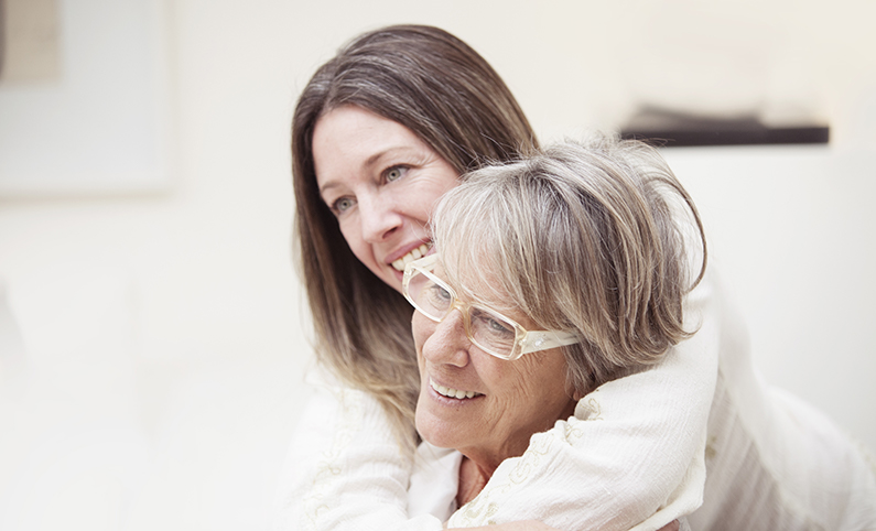
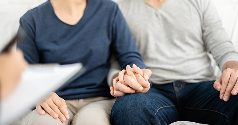
Chances are you or someone you know has a close relative or friend that has been impacted by breast cancer and they may even be receiving breast cancer treatment at one of our clinics. Busting these common myths about breast cancer can help you be informed about what’s real when it comes to this disease.
Breast Cancer Myth 1: Breast cancer only occurs in women.
Though it is true more women are affected by breast cancer, it still can and does occur in men. According to the National Breast Cancer Foundation, over 2,800 men are diagnosed annually with breast cancer. It is important that a man who notices any changes in male breast tissue, including a hard lump underneath the areola or nipple, immediately follow up with his primary care physician.
Breast Cancer Myth 2: A lump in the breast is always cancer.
There are actually many potential causes of a lump in the breast, most of which are not breast cancer. These include the natural changes that occur with aging or any sort of trauma, such as a physical blow, that happened while you were younger. However, it is still important to follow up with your physician if you do notice a lump or change in breast tissue. Being proactive is the best way to detect cancer early, so if it is the reason for your lump, you can be referred to a breast cancer specialist as soon as possible.
Breast Cancer Myth 3: Most breast cancer is genetic.
Only 5 to 10% of breast cancers are thought to result from genetics.It’s not scientifically clear why some women develop breast cancer who are at low risk while some who are at high risk do not. But we do know that most breast cancers are not caused by a hereditary factor. According to the National Breast Cancer Foundation, only 5 to 10% of breast cancers are thought to result from genetics.
There are some risk factors that, if controlled, can reduce the risk of developing breast cancer. This can be done by maintaining a healthy weight, not smoking and keeping daily alcohol consumption to a minimum.
Breast Cancer Myth 4: Breast cancer is contagious.
Very few cancers arise from contagious factors, such as the HPV virus, and breast cancer is not one of them. Breast cancer is the result of cells that have mutated and are growing uncontrollably in breast tissue. It is not contagious and cannot be spread to another person by physical contact, saliva, or any other physical means.
Breast Cancer Myth 5: Deodorant and antiperspirants cause breast cancer.
The National Cancer Institute states that there is no conclusive evidence to show a link between antiperspirant or deodorant use and the development of breast cancer. This means they have reviewed studies that have claimed there is a link and decided there was no sufficient evidence and have also reviewed studies that have failed to demonstrate any link between the two.
Breast Cancer Myth 6: Mammograms can make breast cancer spread.
Some people believe mammograms will cause breast cancer to spread because of the radiation or from the physical compression of the tumor. Neither of these things is true. In fact, mammograms are one of the reasons that deaths from breast cancer have declined, thanks to their ability to detect breast cancer in its early stages when it is more treatable. The patient can be referred to a breast cancer doctor quickly for treatment. While mammograms do deliver a small amount of radiation, the benefit of annual mammograms in women after age 40 outweighs the potential risks. If you still have concerns regarding mammogram screening, be sure to discuss them with your physician, who can help explain why he or she feels mammograms are appropriate for you.
Knowledge Equals Awareness
It’s great that you have taken the time to read about some common breast cancer myths and clear up any misconceptions about breast cancer that you may have had. Many people have heard these myths and have no reason to believe otherwise.
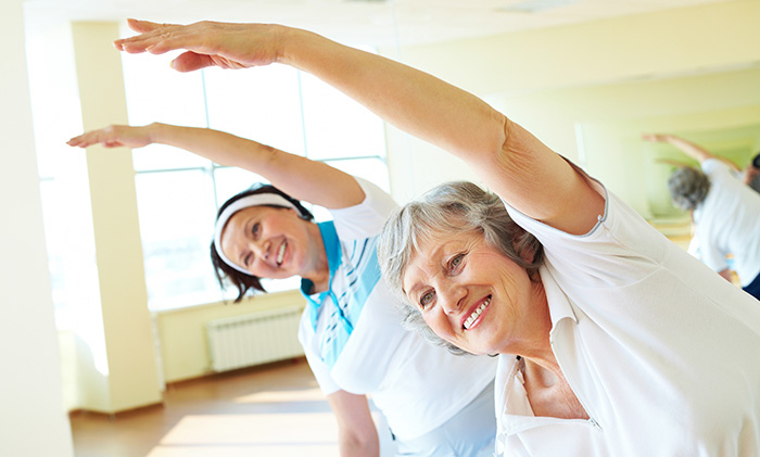
If your oncologist told you there was something safe, free, beneficial and often enjoyable you could do to improve mental and physical health while you undergo active cancer treatment, you might enthusiastically agree right away. Or, you might be skeptical. The good news is that research shows there is such an activity: Exercise!
It’s true that cancer patients grappling with the tolls of radiation and chemotherapy typically don’t make exercise a priority. They can feel physically and mentally exhausted by their cancer treatment regimens. Ironically, if you push through your initial exhaustion, you may discover exercise gives you an abundance of energy, as well as several other benefits. Two studies focusing on breast cancer patients and one on prostate cancer patients showed that exercising during active cancer treatment often yields measurable benefits.
Researchers Study Breast Cancer Patients
It’s well-documented that exercise is beneficial to health. But does it provide extra benefits for individuals undergoing breast cancer treatment? Two studies conclude that for many patients, it does. In the first study, published in the Journal of Community and Supportive Oncology, researchers analyzed the results of 17 clinical trials involving nearly 1,200 breast cancer patients who participated in regular exercise regimens.
Patients participating in aerobic exercises (walking, jogging, swimming, cycling, etc.) experienced marked improvements in quality of life, sleep quality, self-worth, behavior, social well-being, metabolism and aerobic fitness. Patients participating in resistance training (weights, resistance bands, squats, etc.) experienced marked improvements in strength, lean body mass and loss of body fat, and slight improvements in quality of life, fatigue, cognitive function and depression.
Another promising study reported in the Supportive Care and Cancer journal followed 27 breast cancer patients undergoing treatment. The women followed a moderate resistance-training regimen for six months. None of the women developed lymphedema, which is a common side effect of breast cancer treatment.
Researchers Study Prostate Cancer Patients
A third study compared 32 prostate cancer patients participating in a combined aerobic exercise and strength training regimen with a control group of patients not exercising. After three months, the men who exercised during prostate cancer treatment were able to walk much faster than the control group and scored higher on lift-and-carry benchmark tests. The men who exercised also lost, on average, more than four pounds, shed body fat, and reported improvements in quality of life.
Exercise: Little Downside Potentially Huge Upside
Every individual and every cancer patient’s situation is different. Generally, though, experts agree that mild to moderate physical activity offers myriad potential benefits for individuals being treated for cancer. According to the American Cancer Society, exercise may:
Prevent unwanted weight gain
Boost mood and energy
Ward off depression and anxiety
Improve blood flow
Lessen nausea
Lower the risk of heart disease
Prevent muscle atrophy
Restore mobility, including the ability to do day to day activities
Improve patients’ optimism and quality of life
Additionally, multiple studies have concluded that regular exercise may reduce the likelihood of cancer recurrence and increase long-term survival rates.
Considerations Before Beginning an Exercise Regimen
The research is clear: Exercise has little to no downside and potentially a huge upside for patients being treated for cancer. In other words, if you’re a cancer patient and don’t currently exercise, it’s time to talk to your doctor! Taking into account the type and stage of cancer you have, the treatments you’re undergoing, and your physical stamina, strength and fitness level, your doctor should be able to tailor an exercise regimen that will be safe and, hopefully, beneficial to your physical and mental well-being or can refer you to a physical therapist with special expertise in oncology rehab.
Resources from Arizona Oncology
Our team is committed to supporting you throughout your cancer experience. We coordinate and work with many foundations to help support your overall health needs. We’re here to answer questions and connect you with the resources you need, including exercise programs for cancer patients.
(Note: The recommended age to begin colon cancer screening for those at normal risk as been changed to 45.)
I recently turned 50 which meant it was time for my first colonoscopy. Colon cancer is the third leading cause of cancer deaths and generally when caught early, has a good cancer prognosis. Yet, in 2021, 29% of adults age 50 and older had not had a colonoscopy to screen for colon cancer! However, getting your first colonoscopy doesn’t have to be scary, and you can prep to make the procedure go as smoothly as possible.
Here’s what I learned from my first colonoscopy:
Know the colon health of your relatives; if there is a family history of colon cancer, your first colonoscopy might be before the recommended age of 45. Find out more about genetic testing and counseling.
Read and re-read the prep instructions to avoid having the procedure canceled or repeated.
Gather supplies days in advance. The supplies that will help you prepare for your colonoscopy include plush toilet paper, wipes, and a special drink to clean out your colon. A&D ointment can prevent a “sore” rear. Also, choose a variety of clear liquids that you will drink.
Make sure you arrange transportation because you can’t drive yourself home.
Plan your meals. In the days before your procedure, avoid high residue foods like beans, meat, grains, nuts, popcorn, fruits and vegetables which take longer to digest/remove, and can prevent clear visualization of the colon. Avoid foods and liquids that are red, purple or orange. Your prep instructions will include information on all of this.
For your prep, you’ll need to clear your schedule and secure a toilet. Occupy your time and mind; binge-watch a TV series!
None of us experience the prep the same way. Your bowels could start moving right away or take several hours. You’ll experience diarrhea and may have nausea, bloating, and feel cold. It’s important to complete the prep. Your stools should be light yellow and without particles. You’ll get tired, maybe up late and might get a headache from not eating.
Dress comfortably on the day of your procedure, allowing for bloating. When you resume eating, avoid heavy meals and foods that increase gas. Walk frequently to rid the gas. Take a nap.
Your bowels may not be normal for a few days.
The colonoscopy itself was quick and painless. The nicest compliment you can receive for your efforts is that your colon was clean as a whistle.
Not all colon cancers can be prevented but the colonoscopy can save lives; it can find and remove polyps before becoming cancer. On any given day, I would rather have a colonoscopy than to prepare for colon cancer treatments.
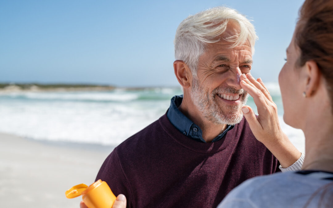
More than two million people in the United States will be diagnosed with skin cancer this year. It is the most common form of cancer in this country, but it is also one of the most preventable. As part of Skin Cancer Awareness Month in May, Arizona Oncology reminds the community about the importance of protecting their skin from the sun and other sources of ultraviolet (UV) radiation.
UV rays are invisible to the naked eye and are more intense in the summer, at higher altitudes, and in areas closer to the equator. Overexposure to ultraviolet radiation from the sun causes sunburn (erythema), skin cancer, premature aging (skin wrinkling), cataracts (gradual clouding of the lens of the eye), immune system suppression, DNA damage and dilated blood vessels.
The most important way for a person to lower his or her risk for skin cancer is to avoid exposure to UV radiation, either from the sun or other sources, such as tanning lamps. The American Cancer Society recommends the following practices for sun safety:
Protect skin with clothing, such as long sleeve shirts and hats with broad brim.
Avoid being outdoors when the ultraviolet light is strongest, particularly between the hours of 10:00 a.m. and 4:00 p.m.
Seek shade
Use sunscreen and lip balm with an SPF of 15 or more on areas of the skin exposed to the sun. Products should be used on hazy or overcast days as well.
Wear wrap-around sunglasses with at least 99 percent UV absorption to provide the best protection for the eyes and the skin around the eyes.
Avoid other sources of UV lights, such as tanning beds and sun lamps.
Protect children from the sun by using the same precautions as adults.
As a community-based cancer care provider throughout Arizona, Arizona Oncology helps cancer patients and their families access a full range of advanced cancer care services in an environment that allows patients to remain close to their homes and their support network of family and friends. Through its affiliation with The US Oncology Network, one of the nation’s largest healthcare services networks dedicated exclusively to cancer treatment and research, Arizona Oncology can quickly bring the latest advances in therapies, research and technology to where patients live. As a result, patients access the best possible treatment with the least amount of disruption to their daily lives.
United In Healing
Arizona Oncology is united in healing with The US Oncology Network. Every day, The Network helps over 2,400 providers deliver value-based, integrated cancer care to patients―close to home. We enable value-based cancer care through an integrated and supportive network that helps comprehensive cancer centers maintain their independence and prosper in today’s evolving healthcare landscape. For more information, visit www.usoncology.com or arizonaoncology.com.
For a man or woman in their childbearing years, a cancer diagnosis can come with a scary thought: will having children be possible? Fortunately, with improvements in treatment and fertility preservation options, having a baby after remission can become a reality for many cancer survivors.
The Risks of Infertility After Cancer
When it comes to whether or not you’re at risk for infertility after cancer, there really is no one-size-fits-all answer. Overall, the chances of remaining fertile depend on a variety of factors including the cancer type, the treatments you received, how your body responded, as well as the original fertility potential.
For both women and men, cancer treatments such as chemotherapy, radiation, and surgery can have a negative effect on fertility. In women, cancer treatment can cause ovarian damage or failure, early menopause, and other reproductive problems that can make it difficult to get pregnant or carry a pregnancy to term. In men, the results of treatment can cause damage to the testes as well as interfere with or destroy sperm production.
The American Cancer Society website has detailed lists of how cancer treatments affect both women and men.
Preserving Fertility Before Treatment
If expanding your family in the future is important to you, speak with your oncologist about how you can preserve your fertility. Prior to beginning cancer treatment, you might want to consider the following options:
Collecting and freezing sperm, eggs, embryos, or ovarian tissue
Gonadal shielding, where unaffected reproductive organs are protected from radiation exposure
Ovarian suppression, which can protect eggs during treatment
Fertility can be damaged by a single cancer therapy session. Additionally, for women, certain methods of fertility preservation are done during certain phases of the menstrual cycle. Because of this, it is important that you talk to your oncologist or a reproductive specialist as soon as possible if you want to preserve your fertility.
Regardless of Your Post-Cancer Fertility, You Can Still Become a Parent
Even if your cancer treatment has caused you to become infertile and you did not take steps or know you could take steps to preserve your fertility before treatment (such as freezing your eggs or sperm) that doesn’t mean you can’t become a parent. Thanks to donor eggs, donor sperm, surrogates who will carry your fertilized embryo to term, adoption, etc. you can still have the family you’ve dreamed of, even if your journey to parenthood is different than you had imagined.
Remember, your cancer care team is your best resource. They can provide you with the most accurate answers to any questions you have regarding the effects of cancer on your fertility, including how long after treatment you should wait before trying to conceive and whether your cancer will be passed onto your child. With support from your cancer care team, friends, and family, you can move forward with hope of becoming a parent.
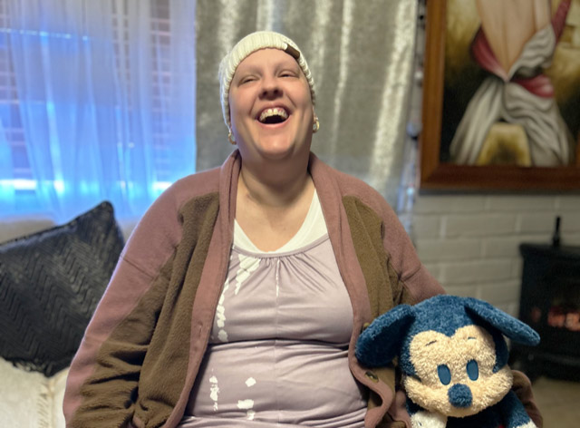
Thirty-eight year old Mandy Hanlon did not expect to get a cancer diagnosis when she arrived at her job as a pharmacy tech on a typical day in August of 2023. She describes what she thought was a sudden onset of nerve pain. Her legs “wouldn’t do what I was telling them to,” and she experienced an abrupt bout of numbness followed by pain.
“The pharmacist saw how much pain I was in and called my dad to pick me up, and we went to Northwest Hospital,” Mandy recalls. “They did a test on me and saw a mass on my back. After tests and more tests, they found out I had stage four bone cancer and tumors in my lungs.”
The diagnosis was Ewing Sarcoma, a very rare form of bone cancer usually found in young people. The doctor she saw first was Arizona Oncology radiation oncologist Dr. Hayden Ansinelli, who reassured her and immediately teamed up with a medical oncologist to reach out into the medical community to ask about treatment options for the disease. “I can’t thank him enough,” Mandy says.
Ewing Sarcoma is a rare form of cancer affecting only one out of one million people in the U.S. Most cases occur in children and adolescents, but there are rare cases that occur in patients over the age of 20. Ewing Sarcoma affects males more than females, often between the age of 10 and 20, and is found mostly in people of European descent. Ewing Sarcoma is also associated with certain genetic predispositions such as neurofibromatosis, Li Fraumani’s Syndrome, and retinoblastoma.
Ewing Sarcoma is potentially curable with a 5-year survival rate around 80% for people whose cancer hasn’t spread. However, for adults the survival rates are lower. Symptoms include bone pain at the tumor site, swelling/redness around the tumor site, fever, weight loss/decreased appetite, fatigue, paralysis and/or incontinence, and nerve compression symptoms such as numbness and tingling.
Initially, Mandy did not want to pursue treatment because of the uncertainty surrounding her disease and prognosis, but her cousin and biggest advocate, Layla, convinced her to try it once to see if it helped. After the first chemo regimen, they saw a reduction in the size of the tumor on her spine, so Mandy decided to move forward with treatment.
The chemotherapy made her quite sick for several weeks, but it was her family and her doctors and nurses who got her through it. During her sometimes 8-hour-long daily chemo treatments, there would be a family member at her side. “I was scared, but all the nurses were amazing. They were my angels,” Mandy says. Mandy had a scare at one point with a complication with her chemo port where her lung filled with blood. “I was so thankful to see my dad when I woke up,” she recalls.
Mandy says her family relationships have been strengthened because of what she’s gone through, especially her relationship with her dad. She adds that the most important thing her caregivers have done for her is to act as her advocate. “There were times when I was on so many pain meds that I couldn’t think or talk, and it was my cousin making sure I understood everything.” Layla also helped her avoid the fees associated with exiting her lease early and made sure she got medical coverage through AHCCCS.
After completing treatment, Mandy reports that while her life has been irrevocably changed by her diagnosis, she’s grateful to be alive. “I will never be able to walk right again, and I am tired all the time, but I am very, very grateful for all the little things, and I try to enjoy the moments more,” she says. On her bucket list is a trip to Disneyland, which she hopes to do in March.
Mandy plans to donate her body to a research facility in the hopes that it will be able to help future Ewing Sarcoma patients, but she has already had a profound impact on those around her. The doctors and nurses at Arizona Oncology have felt it—a sort of gentle groundedness even in the midst of the tumult of emotions cancer brings. When Mandy’s name comes up, they smile. “Wait till you meet her,” they say. And they aren’t wrong. There’s something special about Mandy that everyone who meets her can feel.
“This cancer has changed my life,” Mandy says. “I love myself more now, and I’m so grateful for my family—my dad Michael and stepmother, Alma; they opened a room in their house for me and made me feel comfortable. I am also grateful for my cousin Layla—she was my voice when I was in the hospital, and my Aunt Margie and my sister Shanteal and so many more angels I have around me.”
Mandy encourages others facing a cancer diagnosis to always have faith and look ahead despite the uncertainty and challenges.
“It’s okay,” she says. “Never give up on yourself and keep moving forward.”