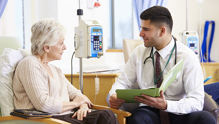
Learning you have cancer is shocking news. After you accept your cancer diagnosis, your main focus is probably, “How can I beat this?” Your oncologist will begin treating you using established, proven treatment protocols based on the specific type of cancer that was diagnosed. In some cases the most commonly effective treatments aren’t working as hoped, and sometimes the cancer returns in other parts of the body. In these cases your oncologist may recommend enrolling in a cancer clinical trial. Should you do this? Find out more about cancer research being done at Arizona Oncology through The US Oncology Network.
Clinical Trials Involve Fine-Tuning FDA-Approved Cancer Treatments
Medical breakthroughs are making new cancer treatments available more often than ever before. Plus, cancer specialists are finding ways to use or combine the already-approved treatments for new uses in cancer treatment. Before these treatments can be made available to everyone they undergo a careful and extensive testing process through clinical trials. There are four primary clinical trial phases.
Phase 1 Clinical Trials: Is the treatment safe?
Research, which leads to human testing, often starts at the test tube level. For cancer, this may include treating cancer cells with the drug(s) of interest.
If a therapeutic response is observed, then the drugs are given to animals. Animal testing provides some insight into possible side effects and generates the starting dose that will be used in the first level of human testing called a Phase I clinical trial. Patients, considered for this cancer treatment, have typically exhausted standard of care options, have an advanced stage of cancer, but have few symptoms from their cancer.
Enrollment in a Phase 1 trial is typically open to patients with various cancers. The first patients are administered the lowest planned dose and observed for side effects over a period of time. If the low dose is tolerated, another group of patients are enrolled at a higher dose level and observed for side effects. Escalating doses of the drug are given to more patients until the maximum tolerated dose is determined. An analysis of the effectiveness against specific cancers is undertaken once the maximum dose is achieved. It is important to understand that not all cancers will respond to any given agent. Only five to ten percent of Phase I agents are effective enough to proceed to the next level.
Phase 2 Clinical Trials: Does the treatment work?
Phase 2 trials have more selective enrollment requirements. The inclusion criteria are based on tumor types that appeared to respond to the Phase 1 trial and often places a restriction on the number of different treatment regimens a patient previously received for their cancer. These patients usually have advanced-stage cancer and are relatively asymptomatic. Many patients are enrolled in a Phase II trial because one of the primary end-points is to prove effectiveness with acceptable side effects. If a reasonable response rate is seen at the Phase II level, the experimental drug or combination can proceed to a Phase 3 trial.
Phase 3: Is the treatment better than what’s available?
Usually, promising drugs undergo FDA approval during phase 3 trials. A Phase III trial usually represents the final destination for a new drug or combination. At this level, the experimental study can be designed for patients with early-stage disease, where a cure is being sought, or more advanced stage cancers where prolongation of life is the goal. Participants are randomly assigned to standard of care chemotherapy or the experimental drug(s). The patients and physicians are blinded to the regimen the patient is receiving. A larger number of patients are enrolled, and the study could take years to complete. The experimental drug(s) can become a new standard of care if they are more effective than the current standard of care, or equivalent but less toxic.
Phase 4: What else can researchers learn about FDA-approved treatments?
In Phase 4 cancer clinical trials, the cancer therapy is tested after it has been approved for a specific use. They are used to collect information about a therapy’s long-term effect on the patient’s quality of life, length of life and any unexpected long-term side effects.
Phase 4 trials may also be used to try new combinations of approved therapies to determine if there are better outcomes.
Most cancer patients who participate in clinical trials in the community setting will join during phases 2 or 3 when researchers are evaluating the effectiveness of new dosages, new combinations of drugs or new uses for existing drugs. Because of this the studies are considered very safe for patients and offer options when other treatments haven’t been effective.
How Do You Participate in a Cancer Research Trial?
You and your oncologist can discuss the potential option of a clinical trial when discussing treatment options. Trials may not be right for all patients. If there is a clinical trial that your doctor and you feel would be right for you, and you agree to participate (you’ll never be added to a trial without your knowledge), you will be carefully monitored throughout the entire process of treatment. This may require more visits than a typical patient would have.
The research team will also contact you regularly after treatment is complete to see how effective it is in the long run.
Who Pays for the Clinical Trial?
Typically there’s no extra cost to the patient for this additional care. The clinical research team will carefully go over this with you.
Many patients who are asked to participate in a clinical trial believe all their care will be paid for through the study. The reality is that only part of their care might be covered. The trial sponsor would provide additional medications as well as funding to cover additional costs considered outside the standard of care practices. Insurance companies are still required to pay for the components of a patient’s care that are considered the standard of care i.e. routine provider visits, laboratory tests, radiographic imaging and prescription medication.
Questions and Concerns About Participating in a Clinical Trial
Because participating in a clinical trial is a different process, there are questions you may have. One concern may be side effects of treatment. Every patient reacts differently, and with new drug combinations or doses there may be new side effects for your oncologist to address. If you participate in a clinical trial but have a poor response, your treatment plan will be changed based on the other options that are available.
If you’re considering participating in a clinical cancer trial, discuss your questions, concerns and expectations with your doctor. Some questions to ask include:
How will my side effects be managed?
What happens if I have a poor response while on the clinical trial?
What’s the purpose of the study?
Is the trial testing new dosages or combinations of a drug that is already FDA-approved, or is it testing a new therapy?
How will I know if I’m qualified to participate?
What will I have to do as a participant? Is there a large time commitment on my part?
How long will the study last?
What type of side effects do you expect?
Will it cost me anything to participate?
Will the researchers tell me the results of the study?
If the treatment is effective for my cancer, can I still get it after the study?
What are the Benefits of Participating in a Clinical Trial for Cancer?
Commonly prescribed medications such as analgesics, antibiotics, and inhalers have gone through the rigorous process of clinical trial investigations. Participation in clinical trials has become an international phenomenon that improves the health of communities. Choosing to enter a clinical trial may provide the benefit of having another opportunity to treat your disease with a non-standard of care option. It is the participation of our friends, neighbors, colleagues, family and strangers that provide us with access to medications and diagnostic tests that have improved the quality of life for all.
If you’re interested in learning about clinical trials appropriate for your specific diagnosis, we encourage you to contact us at Arizona Oncology. We provide Arizona cancer patients access to cutting-edge, innovative cancer clinical trials in a comfortable, convenient setting. Arizona Oncology has locations in and around Northern and Southern Arizona.
In the battle against skin cancer, information is one of the key weapons in your arsenal. With an overwhelming variety of cancer-related articles on the Internet, it’s often hard to tell what’s real and what isn’t.
Let’s debunk the most common skin cancer myths and discuss trusted information sources to help you with further research.
Myth 1: Dark-skinned population doesn’t suffer from skin cancer.
Truth: People suffer from skin cancer regardless of their skin color. According to a study published by the American Cancer Society in 2019, the 5-year relative survival rate for white patients was 94%. For black patients, it was 66%.
The dark-skinned population doesn’t suffer from skin cancer as often as light-skinned people do. However, lack of awareness often leads to late diagnosis, making the survival rate lower. Since the chances of getting skin cancer for dark-skinned individuals is lower, people may not focus on routine screening enough.
This myth is highly dangerous since it can make dark-skinned individuals ignore important health checks. To catch problem areas early, it’s vital to perform a full-body inspection monthly.
Myth 2: After eliminating sun exposure, I can stop worrying about skin cancer.
Truth: Sun exposure is only one of several factors that can lead to skin cancer development. The others include:
Family history – since the chances of developing skin cancer depend on the skin type, if the condition runs in the family, a person may develop it as well. Further research is currently being done to determine which genes may be responsible for increasing the risk of melanoma.
Other UV-light exposure – it’s possible to receive high amounts of UV exposure from tanning beds and occupational equipment. It’s important to understand that the culprit isn’t the sun. It’s UV rays. Anything producing UV rays can be a potential hazard.
It’s worth stressing that a tanning bed isn’t less dangerous than sun exposure. Your skin receives the exact same UV rays.
Truth: According to the Skin Cancer Foundation, the difference between SPF 30 and SPF 100 is very slight. Raising the sun protection factor is often a marketing trick, which gives you a false sense of protection.
In reality, sunscreens with SPF 30 block out 97% of harmful UV rays. SPF 50 protects you against 98% of UV rays. SPF 100 can absorb up to 99% of UV rays.
This myth is highly dangerous because people feel falsely protected against the sun and tend to stay outdoors longer. Even the highest sun protection factor can’t keep you 100% safe. The best way to avoid skin cancer is to stay out of the sun or wear protective clothing.
Myth 4: People who rarely get sunburned are at a lower risk of getting cancer.
Truth: A healthy suntan doesn’t exist. Even if you tan easily and never get sunburned, you can get skin cancer.
There is some truth in the above-mentioned myth. Skin type is a major component of skin cancer risks. According to the World Health Organization, people with fair skin are at a bigger risk for skin cancer.
However, excessive sun exposure damages your skin even if you don’t feel it. No matter what skin type you have, the risk of acquiring skin cancer is always present.
This myth is highly dangerous. People who suntan easily and don’t get burned may be spending more time in the sun since they don’t feel its effect. This high degree of carelessness can lead to serious consequences.
Even if you don’t feel sunburned, you are at risk of getting skin cancer.
Myth 5: If you’re young, you don’t need to worry about skin cancer. It affects older people only.
According to the Centers for Disease Control and Prevention (CDC), the majority of skin cancer sufferers fall into the 80-84 age group. However, people as young as 15 can acquire skin cancer.
One of the reasons why older people are at a higher risk of getting skin cancer is the cumulative effect of sun exposure. Meanwhile, people with weak immune systems and other risk factors may not need excessive sun exposure to suffer from its consequences.
Myth 6: Sun exposure is less dangerous when the sky is cloudy.
Truth: When the day is cloudy, the sun doesn’t go anywhere. It continues to shine above the clouds, sending harmful UV rays to the surface. While cloud cover may reduce sun exposure, it doesn’t eliminate it entirely. That’s why it’s important to maintain the same level of sun protection when it’s cloudy outside.
When the sky is fully covered by clouds, sun exposure is lower. However, broken and scattered clouds don’t provide much protection.
Myth 7: Skin cancer appears only on those parts of the body that were exposed to the sun.
Truth: Skin cancer can appear on any part of the body, including feet soles, palms, underneath the fingernails/toenails, and on the genitals. That’s why it’s important to inspect the entire body for the signs of skin cancer regularly.
Trusted Sources of Information
It’s tempting to believe in some of the common myths surrounding skin cancer. However, when it comes to your health, it’s important to use trusted sources. If you want to conduct online research, take advantage of the following trusted websites:
More people than ever are working from home. Some of the perks that come with this working from home include flexible hours, spending more time with family, and leaving your commute behind. Unfortunately, you could also be exposing yourself to additional skin cancer risks that you don’t normally face. Watch for these risks and use practical tips to prevent extra sun exposure while working from home.
1. You’re More Relaxed
Relaxing definitely doesn’t put you at a higher risk of cancer. However, as you make changes to your daily routine, you may relax certain daily habits that go along with your work schedule. For many people, the daily application of sunscreen comes during the process of getting ready for work. Changing that schedule could mean forgetting this essential protection. With practically 300 sunny days in Arizona each year, letting your guard down means letting skin cancer risks in.
Maintaining Important Health Regimens
There’s no denying that a flexible schedule is one of the biggest benefits of working from home. Still, your health should never take a back seat. No matter what your daily routine looks like, make sunscreen application a priority. Associate sunscreen with another morning habit you never forget like taking medication or brushing your teeth.
2. Working Outside the Office
Even if your workplace promotes the most flexible working style in the area, most work hours are spent indoors. The flexibility of working from home may inspire you to take breaks outside, take walks during the day for exercise, or work outdoors on your deck or patio. The sun’s rays are strongest between 10:00 AM and 4:00 PM. Many workers are accustomed to spending this time inside the workplace. When you’re not spending these hours within the confines of a work building, you are likely accumulating extra hours of sun exposure.
Staying Safe Out of the Office
Learning the facts about skin cancer and the most effective ways to prevent it is key to avoiding dangerous habits. Chart the unexpected time you’re spending outdoors and how often you apply sunscreen. For sunscreen to be effective, it must be used correctly. Use these tips to get the best defense with sunscreen.
Apply sunscreen to dry skin 15 to 30 minutes before going outdoors.
Use sunscreen with an SPF of 30 or higher.
Get a new bottle of sunscreen each year. The ingredients in sunscreen break down over time and become less effective.
Use a teaspoon for your face and neck and enough to fill a shot glass for the rest of your body. When in doubt, add more.
Reapply sunscreen every 90 minutes while outdoors. Always reapply sunscreen after swimming or after excessive sweating.
Don’t skip those easily forgotten places like your ears, the back of your neck, and tops of your feet.
Use sunscreen even on cloudy days.
3. Spending Time Outside With the Kids
You’re not at work and the kids aren’t at school. Suddenly, your priorities shift and everyone is outdoors in the Arizona sunshine. There is an abundance of benefits to spending time outside like exercise and fresh air, but these extra hours outdoors add up when it comes to sun exposure. An hour or two of unplanned time outdoors can lead to dangerous sunburns and skin damage. Even a few sunburns can increase your children’s risk of skin cancer.
Safe Skin Habits for You and Your Children
Safe health habits start early and remain consistent. The same way you stop to wash your hands before eating or put on a jacket when there’s a chill in the air, you should stop to put on sunscreen before going outdoors. Sunscreen should be applied to dry skin at least 15 minutes before going outside and reapplied every two hours or immediately after swimming or sweating. Additional ways to stay safe include spending time in the shade, wearing protective clothing, and wearing a hat and sunglasses.
4. Lack of Task Lighting
If you’ve recently moved from a brightly lit workplace, your new “office space” might be right next to a window to take advantage of natural light. While natural light is often preferable to harsh fluorescent lighting, dangerous UV rays can penetrate glass to cause damage to your skin and put you at a higher risk for skin cancer.
Safe Use of Natural Lighting
Eliminating natural light completely would be a shameful waste of Arizona’s natural resources. Glass does provide some protection from the sun’s rays, so you don’t need to board up the windows. Applying sunscreen daily and sitting by a window that doesn’t provide direct sunlight is likely enough to protect your skin indoors. If your only home office option is near a window that sees an abundance of direct lighting, consider UV blocking window film for added protection.
5. No More Uniforms
Whether your workplace requires a standard uniform, business attire, or certain dress code rules, that wardrobe goes out the window when you work from home. Your personal wardrobe reflects your sense of style and, more often than not, the weather outdoors. Unfortunately, this often means that your casual clothing leaves more skin exposed to the elements than the clothes you typically wear to work.
A Uniform for Working Outdoors
You don’t need a uniform to work at home, but a type of dress code is necessary when you’re spending time outdoors. Your best line of defense against the sun’s harmful rays is a physical barrier of clothing. Tightly woven dark fabrics provide the most protection. Additional accessories can add an extra layer of protection. A wide brim hat and sunglasses provide a shield for delicate facial skin.
Working from home means a change in your routine. While you’re getting used to these changes, it’s important to take precautions that will protect your skin from the sun’s dangerous UV rays. Skin cancer is the most common cancer diagnosis and also the most preventable. Forming habits to protect your skin is the best way to prevent a skin cancer diagnosis.
Metastatic breast cancer, which may also be referred to as Stage IV breast cancer, indicates that cancer has spread from the breast tissue and the nearby lymph nodes to other organs in the body, most commonly the bones, lungs, liver or brain. Any type of breast cancer (estrogen-positive, HER2-positive, etc.) can metastasize (spread) to other areas of the body.
When a tumor is found outside of the breast, it’s made up of breast cancer cells. For example, if you have a tumor in the lungs that is metastasized breast cancer, it contains breast cancer cells, not lung cancer cells. These cells may no longer react to the treatments given in the past, meaning that new cancer therapies may be necessary.
The sooner you can detect cancer that has spread to other areas of the body, the easier it will be to contain and treat. That means it’s important to know some of the signs of metastatic breast cancer.
What Are the Signs of Metastatic Breast Cancer?
You will usually show some of these signs before receiving confirmation that the breast cancer has spread. However, you may not associate these symptoms with metastatic breast cancer when only one or two appear. Be sure to talk to your oncologist if you notice any of the following:
Consistent back, joint, or bone pain
Confusion
Severe headache
Difficulties urinating. This can include incontinence or not being able to urinate at all. This happens when the cancer pinches the nerves in your back.
Numbness or weakness anywhere in your body
Difficulty breathing
Chest pain
Bloating, pain, or tenderness in the abdominal region
A constant dry cough
Vision issues, such as blurry vision, loss of vision, or double vision
Shortness of breath
Chest pain
Loss of appetite
Balance issues
Signs of jaundice, such as a yellow tinge to your skin or the whites of your eyes
Consistent nausea, vomiting, or weight loss
Seizures
These symptoms can also develop from other medical issues. After talking to your oncologist, he or she may ask for some tests to be run to see if there are breast cancer cells in other areas of the body.
How Does Your Oncologist Determine Metastatic Breast Cancer?
If you’re seeing a different cancer care provider than you did on your last round of treatment, it’s a good idea to bring all of your medical records with you from the previous oncologist. This will help your current cancer specialist understand the type of cancer, its stage, and what was done to treat it. This information can have an impact on what steps are taken next.
After meeting with the oncologist, there are likely some tests to be run to determine what may need to be done next. Here are some common tests for breast cancer detection:
Blood tests. This can include tumor markers in some patients.
Whole-body bone scan and this can include X-rays of specific bones
CT scan of the chest, abdomen, pelvis, and/or brain
Bronchoscopy for patients with a constant cough or trouble breathing
Spinal tap to remove fluid from around the spinal cord
Pleural tap checks the fluid between the chest wall and lungs
X-ray or ultrasound of the abdomen or chest
Your doctor may start out by ordering a couple of tests and based on the results may request further testing. This is so they can get a clear picture of how extensive the cancer has spread throughout the body. This also helps your oncologist come up with the treatment plan they feel will work best for you.
Treatments for Metastatic Breast Cancer
Once you have confirmation of metastatic breast cancer, and your oncologists have the information they need, a treatment plan can be created. In some cases there is a combination of treatments required. One or more of the following may be included in your treatment plan for metastatic breast cancer:
Hormone therapies: The most common approach and treatment for metastatic estrogen or progesterone receptor-positive breast cancer is hormone therapy. You may have already used one or more of these in your previous treatments. Based on your history, the oncologist may recommend a new hormone therapy. The most common hormone therapies are:
Tamoxifen: This treatment blocks estrogen in both pre and postmenopausal women. It’s used primarily in young women who haven’t had hormone therapy before, and it’s designed to contain and shrink the tumors.
Fulvestrant: After attaching to the estrogen receptors, this drug changes the shape of the receptors and degrades it preventing further tumor growth. This should slow the growth of cancer cells.
Aromatase inhibitors: These hormones stop the actions of the enzyme aromatase, which is responsible for the conversion of androgens to estrogens. The hope is that this will lower levels of circulating estrogen and stop the growth of cancer cells.
There are other hormone therapies being introduced for advanced breast cancer. Talk to your oncologist about what is available if you have used these other hormone therapies already.
Chemotherapy drugs: If you’ve already battled breast cancer, you may find some drugs you’re familiar with while others are used more often for metastatic breast cancer. This is the most common approach for metastatic breast cancer that is :
Hormone-receptor negative
Hormone-receptor positive, but doesn’t respond to hormone therapies
HER2-positive, in combination with a targeted therapy
Chemotherapy tends to work quicker than hormone therapy, which may also be a consideration for whether it’s used right away.
Targeted therapies: Most commonly used for HER2 positive breast cancers, this category of drug targets the HER2 protein on the cells which fuels cancer cell development.
Immunotherapies: These drugs, which help your immune system fight the cancer, are less commonly used for breast cancer, but may be used in combination with other treatments.
Surgery: Sometimes surgery to remove a tumor that has developed in another area of the body is necessary to avoid further growth. The location of the tumor dictates if surgery is a possibility.
If you’ve been previously diagnosed with breast cancer, it’s critical that you attend your regularly scheduled check-ups, even years later. At the first signs of metastatic breast cancer, you need to contact your oncologist.
At Arizona Oncology, we understand your apprehension, and we’re ready to help you at each step of the process.
Cancer treatments such as chemotherapy and radiation are designed to kill fast-growing cancer cells. These powerful drugs travel throughout the body and may affect healthy, normal fast-growing cells as well. The type of long-term side effects cancer survivors face and the severity differs between individuals. Some may experience minimal long-term effects, while others may experience moderate to severe long-term effects.
There are many different types of cancer and many different types of treatment. This combined with the fact that everyone’s body responds differently makes the possibilities of long-term side effects from cancer treatment endless. Let’s take a look at the most common long-term side effects:
Side-effects from surgery
Dental and oral issues
Heart conditions
High blood pressure
Pulmonary conditions
Side-effects to the endocrine system
Impaired brain function/chemo brain
Hair loss
Secondary cancer
Decreased bone strength
Emotional problems
Long-Term Side Effects Related to Surgery
Long-term surgical side effects(1) will depend on the type of cancer you were diagnosed with and the location in the body you had surgery. The following are a few examples of side effects from surgery. Scarring at the surgical site is a common long-term effect as well as chronic pain. Lymphedema, which is painful swelling from fluid build-up is the result of having lymph nodes removed. Survivors who had a limb amputation may experience phantom pain. If your spleen was removed due to Hodgkin’s Lymphoma, you might be at a higher risk for developing infections.
Dental and Oral Issues
Tissues in the mouth are among the fastest-growing tissues in the body. Since cancer treatments attack fast-growing tissues it can cause long-term negative dental and oral side-effects such as:
Cavities
Tooth decay
Dry mouth
Gum disease
Loss of taste
It is important to see your dentist if you experience any oral issues after cancer treatment.
Heart Conditions
Chemotherapy and radiation cancer treatment to the chest increases the risk of long-term side effects on the heart. Congestive heart failure, abnormal heart rhythms, and hardened and narrow arteries (coronary artery disease) are the most common side effects, although they may not appear for months or even years after cancer treatment is complete. There is also an increased risk of developing heart issues when taking certain medications as a part of your cancer treatment.
High Blood Pressure
Certain medications used as a part of cancer treatment can cause high blood pressure in patients. These include Bevacizumab, Sorafenib, and Sunitinib.(1) For many patients, blood pressure returns to normal once you are no longer taking the medication. However, for others, it is a long-term side effect that must be managed with medications, diet, exercise, or a combination of treatments to keep your blood pressure under control.
Pulmonary Conditions
Those who received either chemo or radiation to the chest are more likely to experience damage to the lungs. Survivors who received both chemo and radiation are an even higher risk of long-term pulmonary side effects due to cancer treatment. Side-effects include:
A decrease in pulmonary function
Difficulty breathing
Inflammation and thickening of the lining of the lungs
Long-Term Side Effects to the Endocrine System
Cancer treatments may cause long-term side effects to the endocrine system due to the chemotherapy or radiation causing a change in the level of hormones for both men and women. For women, this could mean menopausal symptoms in which your body stops menstruating. For younger women, menstruation may come back at some point after cancer treatments are finished. This is not always the case.
For both men and women, the change in hormones due to treatment can cause infertility, changes in your sex drive, and hot flashes.
Chemotherapy and radiation can cause survivors to have long-term trouble with memory, processing information, and staying focused on tasks at hand. Those who received cancer treatment to the head are at a higher risk for developing long-term cognitive issues. This is commonly referred to as chemo brain. While chemo brain is usually worse during treatment, it is known to last after treatment is completed-sometimes for a few years.
Fatigue
Cancer treatment is powerful and can not only leave the patient feeling tired during treatment but for months and years afterward. This is one of the most common long-term side effects experienced by cancer survivors.
Long-Term Bone Issues
Certain cancer treatments may cause thinning of the bones, which is also called osteoporosis. Survivors may also experience joint pain as well. These side-effects can be made worse if the individual is not physically active.
Hair Loss
Hair loss is a common side-effect of cancer treatment. For most people, their hair begins to grow back after treatment. For others, it may not grow back, it may be thinner, and it may come back a completely different color or texture.
Secondary cancers, although not common do happen. It is important to understand that a secondary cancer is not a recurrence of the original type of cancer. It is an altogether different and unrelated cancer that occurs in a different part of the body. For instance, let’s say you successfully completed breast cancer treatment and years down the road are diagnosed with colon cancer. Colon cancer is a secondary cancer.
Certain chemotherapy and radiation treatments for cancer have the potential to increase the chances that an individual will develop a secondary cancer down the road. The younger an individual is when they undergo cancer treatment, the higher the risk of developing a secondary cancer later in life.
Emotional Difficulties
It is very common that cancer survivors experience a range of emotions from relieved and happy to sad, angry, depressed, helpless, and everything in between. From the moment you are diagnosed to when you receive your last treatment and beyond is a roller coaster of emotion for both the survivor and their families. Talking with someone can help survivors work through their emotional difficulties.
Managing Long-Term Side Effects Due to Cancer Treatment
Cancer treatment takes a toll on the mind and body, that is not a secret. It stands to reason that it takes time to begin feeling like yourself again. Arizona Oncology understands that the journey does not end when treatment ends. There are both the physical and emotional side-effects of being faced with and surviving a life-threatening disease. You do not have to go it alone. Our staff offers supportive care to help you manage your long term side effects and adjust to a new normal in life!
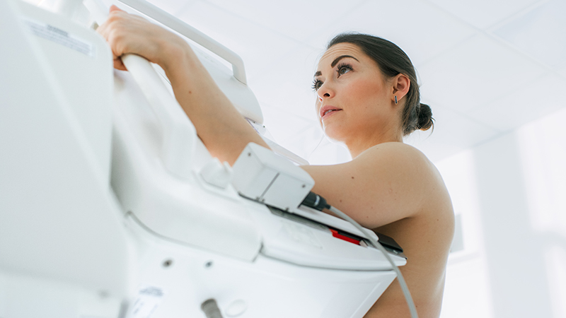
If you are preparing for or anticipating having to schedule your first mammogram, you may be wondering what to expect. A mammogram is a non-invasive diagnostic scan essential for early detection of breast cancer. It can be an inexpensive and highly effective method for reducing breast cancer risks. Having regular mammograms can be critical for those with a higher risk level or history of family breast cancer of any age.
Today, we’ll provide a guide to preparing for your first mammogram and outline what you can expect during your first screening.
When Should You Have Your First Mammogram?
Many experts suggest annual mammograms are ideal for women within the 45-54 age range. Annual screenings are often recommended and can significantly reduce the risks of death as a result. In fact, research suggests regular mammograms reduce breast cancer death risks by 14% for women within 50 and 60 years old. Those percentages increase with age; up to 33% for those 60 to 70 years old. Some women may begin mammogram routines earlier in life, depending on individual health conditions, family history of breast abnormalities, or the discovery of a lump. Your health care provider can help you determine when it’s best to begin your annual mammogram screenings.
Scheduling & Choosing a Facility
Your health care team or primary care physician may recommend a facility for your first mammogram. If you don’t have a recommendation to guide you, consider finding an area location that routinely performs mammograms. As you prepare for your first mammogram, you’ll want to choose a site that can perform your annual mammograms in the years to come. Staying with one provider can make it easier to compare results year after year.
Preparing for Paperwork & Forms
You will want to be mindful of your specific insurance carrier information ahead of time. Most insurance providers cover mammogram procedures as a standard, preventative visit. However, it’s best to review your plan to understand what coverages apply to you. You will be asked to present your card at the time of your appointment. It is a common suggestion that you arrive to the appointment a little early, should you have insurance or provider related questions. Being early can also allocate enough time to complete any new patient, or medical history forms the facility may require.
Preparing for the Mammogram
When you are called back, you can expect to be situated in a private room. The technician will provide you with a waist-up gown for you to wear. In preparation before you go, it’s best to make sure you’re not wearing any jewelry or deodorant. It’s also recommended that you not be wearing lotions or skincare products. The types of products can affect the imaging. If you arrive to your appointment having forgotten to abstain from these products, the technician can provide you with a warm cloth or towelette to remove them.
What to Expect During the Imaging Process
You can expect the procedure itself to be relatively brief. The technician will position the machine, and you, in order to take pictures of the breast. Once you are strategically positioned, you’ll be asked to remain completely still and maybe to hold your breath for a few brief seconds during the picture-taking process. You can expect a few different positions for each breast. The technician may leave the room to digitally present the images to the on-staff Radiologist for quality review. If any of the pictures are obscure, the team may have you repositioned for a retake or two. Once they are confident the images are clear, you’ll be free to get dressed and leave.
Are Mammograms Painful?
While you can expect mild discomfort during the mammogram process, it’s relatively painless. Your breast will be positioned on a tray that is adjusted to accommodate your height. A top-level tray will be lowered to hold your breast in place. So, while it can be uncomfortable for a few moments, you won’t encounter any significant pain. As an added tip, try to avoid scheduling your mammogram during the first week of or week prior to starting your period. Because your breasts can be sensitive during this time, the position requirements of a mammogram can be slightly more painful.
Most routine mammogram results will be delivered to you by mail or phone and within ten days. You may be notified sooner, depending on the facility. Upon review of the images, the Radiologist may want additional scans, ultrasounds, or x-rays. Don’t be alarmed if you receive notice to return for another round of images. It may mean there is an unclear area, that one of these other methods of imaging can portray better. Your referring physician can be of assistance during this follow-up and waiting period as well, should you have questions about follow up scan requests. Just know that callbacks can be common, and most prove not to represent breast cancer.
Getting Started on Your Path of Mammogram Routine Screening
It can be scary for some women to consider embarking on the annual mammogram routine. There may always be a subtle concern or anxiety that mammograms will uncover breast cancers. The good news is, screening often results in normal findings. For those instances when abnormalities are present, the early detection alone can be life-saving. If you’re approaching the age of 45, it’s a good idea to add mammograms to your roster of health-promoting and preventative visits. If you’re younger, but have concerns with family history or a recently discovered lump, ask your primary care physician for guidance or referral to an area mammogram facility.
Consider mammograms as a necessary and diagnostic tool that helps better arm women to combat the risk of breast cancer. Images can provide valuable information about your current breast health. They can also serve as a timeline of changing breast health in the years to come. Being able to identify a mass early, or changes to breast tissue can help ensure your best chances of recovery, survival, and health preservation overall.